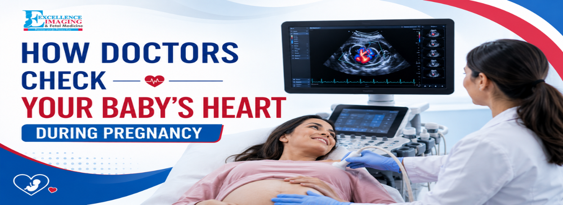
Hearing your baby’s heartbeat for the first time is a moment you will never forget. It is a beautiful, rapid sound that brings instant peace of mind to expectant parents.
But when doctors look at the monitor, they are checking far beyond just the sound. Monitoring how the heart forms is a routine, step-by-step process that happens throughout your entire pregnancy journey.
By understanding how a specialized diagnostic centre checks your little one, you can walk into every prenatal appointment feeling completely relaxed and informed.
Specialized Diagnostic Centre
In the early weeks of pregnancy, the focus is simply on confirming that everything is starting exactly as it should. Because the embryo is still incredibly small, a highly detailed fetal heart scan isn't possible yet. Instead, doctors use simple tools to look for baseline signs of life.
Early Ultrasounds
During your first checkup, usually around weeks 6 to 9, a diagnostic centre will perform an early pregnancy heart scan. The sonographer looks for a tiny, rhythmic flickering on the screen, which shows that the tiny cardiac tube has started pumping.
Handheld Doppler Checks
As you near the end of your first trimester, your doctor will start using a small handheld Doppler device during routine visits. This device uses safe sound waves to amplify the audio of the heart beat so you can hear it out loud in the room.
Second Trimester: The Mid-Pregnancy Anatomy Scan (Weeks 13 – 27)
Between weeks 18 and 22, you will have your standard mid-pregnancy anatomy ultrasound. This is a very important milestone because it includes a detailed baby heart scan to check the actual structure of the heart.
Checking the Four Chambers
The most important part of this routine fetal heart ultrasound is looking at the "four-chamber view." The sonographer pauses the screen to make sure the internal structure is shaped correctly.
- Size and Balance: They check that the left and right sides of the heart are perfectly symmetrical.
- The Central Wall: They look closely at the wall separating the chambers to ensure it is solid and whole.
- Working Valves: They watch the tiny internal valves open and close naturally to keep blood moving.
Tracking the Main Blood Vessels
During this same fetal cardiac scan, the doctor also checks the main pathways leading away from the heart. They need to see that the primary blood vessels are connected to the correct chambers so that oxygen travels smoothly to the rest of the baby's body.
Specialized Scans: When Doctors Need a Closer Look (Weeks 28 – Birth)
For most parents, the routine mid-pregnancy ultrasound gives all the answers needed.
However, there are times when a doctor might want a more focused view.
If the baby was in a tricky position during the regular check, or if there is a family history of
cardiac issues, your doctor might suggest a deeper look.
Why Is a Deeper Scan Suggested? A dedicated fetal cardiac ultrasound is a normal precautionary step if the mother has a condition like diabetes, or if a routine screening shows a slight irregularity in the rhythm.
This is exactly when fetal echocardiography is recommended It does not mean something is wrong. Most of the time, it is simply done to give parents ultimate peace of mind.
How an Advanced Fetal Echo Works
If you are referred for a specialized fetal echocardiography scan, the actual experience is incredibly simple and completely painless for the mother. It feels just like a regular ultrasound.
Color Flow Mapping
During this specific fetal heart ultrasound, the specialist turns on a feature called color Doppler.
This turns the black-and-white screen into bright red and blue colors.
These colors show the exact path and speed of the blood flowing through the tiny valves. It
helps the doctor confirm that everything is circulating perfectly.
Planning for a Healthy Birth
The main reason doctors perform these detailed checks is to screen early for any congenital
heart defects in baby growth.
If a heart defect in a baby is caught early on a scan, it allows families and doctors to plan
beautifully for delivery day. A specialized imaging centre provides the clear pictures needed so
that pediatric experts can be right there to support your baby the moment they are born.
Choosing a Trusted Team for Your Prenatal Care
When it comes to your pregnancy, having advanced technology and experienced eyes reading your scans makes all the difference in the world. At Excellence Imaging Center, we combine gentle, compassionate care with the latest imaging tools. Whether you are coming in for a routine baby heart ultrasound or a detailed fetal echocardiography scan, our centre is here to keep you safe, informed, and completely reassured at every step.
Frequently Asked Questions (FAQs)
Question - Is a baby heart scan completely safe for my pregnancy?
Answer - Yes, it is entirely safe. Every routine checkup and advanced fetal heart ultrasound
uses simple sound waves to see the baby. There is absolutely no radiation involved, making it
perfectly safe for both mom and baby.
Question - What makes a fetal echo different from a regular ultrasound?
Answer - A routine anatomy scan looks at the baby’s whole body, including a quick look at the heart. A specialized fetal echo test focuses 100% of the time on the intricate details, valves, and pumping actions inside the cardiac system.
Question - Can a prenatal scan find every single heart issue?
Answer - While a detailed baby heart scan is incredibly accurate at showing structural shapes, a few minor conditions—like a very tiny hole or a minor valve change—might only be discovered after the baby is born and starts breathing on their own.
Question - Do I need to do anything special to prepare for the test?
Answer - No special preparation is needed for a heart scan during pregnancy. Unlike early pregnancy scans, you do not need a full bladder for this test. Just wear comfortable clothing so the sonographer can easily reach your stomach.
Question - Will my doctor check for structural issues at every visit?
Answer - No, a highly focused baby heart ultrasound checking for congenital heart defects in baby development is usually done during your main second-trimester appointments. At regular weekly checkups, your doctor will simply use a quick Doppler to listen to the rhythm and speed of the beat.